Abstract
Purpose: Overall, this study plans to illustrate the ability of EO sclerotherapy as an alternative to surgery, with greater efficacy and reduced incidence and severity of risks.
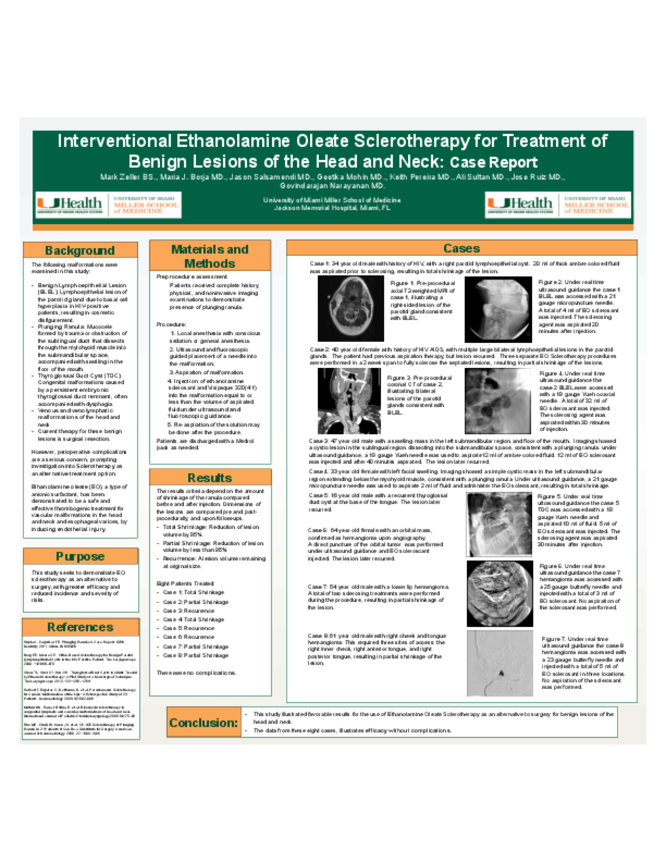
Background: The malformations examined in this study include plunging ranulas, benign lymphoepithelial lesions (BLEL), a thyroglossal duct cyst (TDC), and venous and venolymphatic malformations of the head and neck. A plunging ranula is a mucocele formed by trauma or obstruction of the sublingual duct that dissects through the mylohyoid muscle into the submandibular space, accompanied with swelling in the floor of the mouth. BLEL are lymphoepithelial lesions of the parotid glands due to basal cell hyperplasia in HIV-positive patients, resulting in cosmetic disfigurement. TDC are congenital malformations caused by a persistent embryonic thyroglossal duct remnant, often accompanied with dysphagia. Venous malformations occur when vessels anastomose irregularly and dissect the normal components of the surrounding tissue. Current therapy for these benign lesions is surgical resection. However, perioperative complications are a serious concern, prompting investigation into sclerotherapy, which has been reported as an effective treatment option.
Methods: In our study, ethanolamine oleate (EO), a type of anionic surfactant, is used as the sclerosing agent. EO has been demonstrated to be a safe and effective thrombogenic treatment for vascular malformations in the head and neck and esophageal varices, by inducing endothelial injury. EO is mixed with radiopaque Iodixanol, a contrast agent, in a 8:2 ratio to demonstrate the distribution of the sclerosant during injection.
A total of eight patients participated in this ongoing study. Two patients presented with plunging ranulas, two patients presented with BLELs, one patient presented with a TDC, one patient presented with lower lip venous malformation, one patient presented with a venolymphatic malformation of the neck, and one patient presented with a venolymphatic malformation of the head and neck. Patients receive complete history, physical, and noninvasive imaging examinations to demonstrate presence of the malformation. The malformation is accessed under real-time ultrasound guidance. Upon aspiration of the malformation contents, an equal volume of the EO and Iodixanol mixture is injected into the malformation with fluoroscopic guidance. Patients undergo follow-up ultrasound after 4 weeks, and then as necessary depending on signs of recurrence.
Results: Dimensions of the lesions are compared pre and post-procedurally, and upon follow-ups. A reduction of lesion volume by 95% constitutes “total shrinkage”. A reduction of lesion volume by less than 95% constitutes “partial shrinkage”. A lesion volume remaining at original size constitutes recurrence. An increase in lesion volume past original size constitutes progression.
Of the eight patients treated with this therapy, three patients experienced total shrinkage (ranula, BLEL, and neck malformation), four patients experienced partial shrinkage (ranula, BLEL, lower lip, and head and neck malformation), and one patient experienced recurrence (thyoglossal duct cyst).
Conclusion: This study illustrated favorable results for the use of Ethanolamine Oleate Sclerotherapy as an alternative to surgery for benign lesions of the head and neck.
The data from these eight cases, illustrates efficacy without complications.