Abstract
Introduction: The spontaneous rupture of a common iliac artery (CIA) is rare and limited, even more so in isolation of an abdominal aortic aneurysm (AAA) with the internal and external iliac arteries being the most common locations. Etiologic factors associated with this unusual entity include trauma, atherosclerosis, cystic medial necrosis, high level sporting exercise and connective tissue disorders (CTD’s) such as Marfan Syndrome, 1-antitrypsin deficiency, Ehlers-Danlos and Fibromuscular Dysplasia. Rupture of a CIA is associated with a higher mortality rate with approximately 30% following open repair. Endovascular stent grafting for emergent repair has lowered perioperative mortality rates. The risk of rupture is greater when the site of aneurysm or dissection is at the CIA.
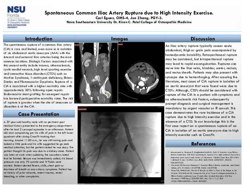
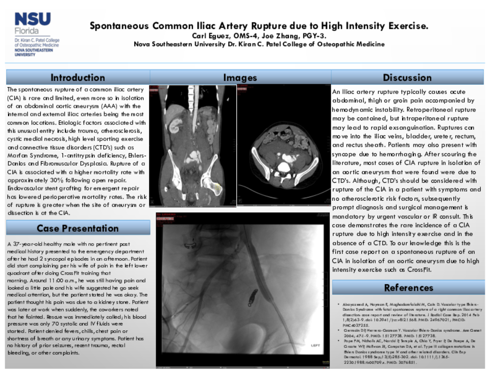
Case Description: A 37-year-old healthy male with no pertinent past medical history presented to the emergency department after he had 2 syncopal episodes in an afternoon. Patient did start complaining per his wife of pain in the left lower quadrant after doing CrossFit training that morning. Around 11:00 a.m., he was still having pain and looked a little pale and his wife suggested he go seek medical attention, but the patient stated he was okay. The patient thought his pain was due to a kidney stone. Patient was later at work when suddenly, the coworkers noted that he fainted. Rescue was immediately called; his blood pressure was only 70 systolic and IV fluids were started. Patient denied fevers, chills, chest pain or shortness of breath or any urinary symptoms. Patient has no history of prior seizures, recent trauma, rectal bleeding, or other complaints. CT abdomen with contrast was done and indicated mild ectasia of distal left common iliac artery measuring 10 mm in diameter. Massive contrast extravasation from the left common iliac artery with associated large amount of hemorrhage throughout the retroperitoneum in the abdomen and pelvis. Retroperitoneal hematoma measured roughly 9.5 x 19 cm in anterior and posterior and transverse dimensions and the inferior vena cava was collapsed. Expeditiously, the hematoma was evacuated, and a stent was placed by an Interventional Radiologist.
Discussion: An Iliac artery rupture typically causes acute abdominal, thigh or groin pain accompanied by hemodynamic instability. Retroperitoneal rupture may be contained, but intraperitoneal rupture may lead to rapid exsanguination. Ruptures can move into the iliac veins, bladder, ureter, rectum, and rectus sheath. Patients may also present with syncope due to hemorrhaging. After scouring the literature, most cases of CIA rupture in isolation of an aortic aneurysm that were found were due to CTD’s. Although, CTD’s should be considered with rupture of the CIA in a patient with symptoms and no atherosclerotic risk factors, subsequently prompt diagnosis and surgical management is mandatory by urgent vascular or IR consult. This case demonstrates the rare incidence of a CIA rupture due to high intensity exercise and in the absence of a CTD. To our knowledge this is the first case report on a spontaneous rupture of an CIA in isolation of an aortic aneurysm due to high intensity exercise such as CrossFit.