Abstract
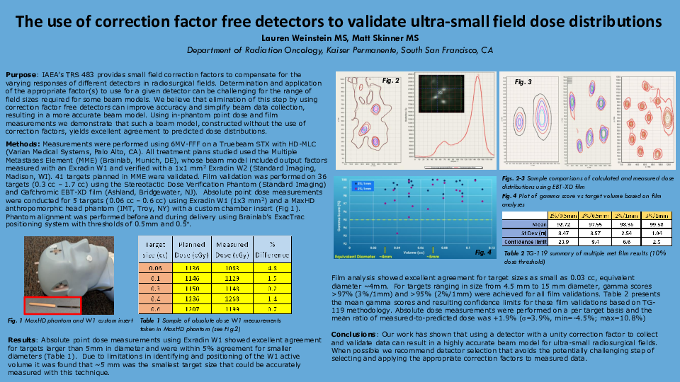
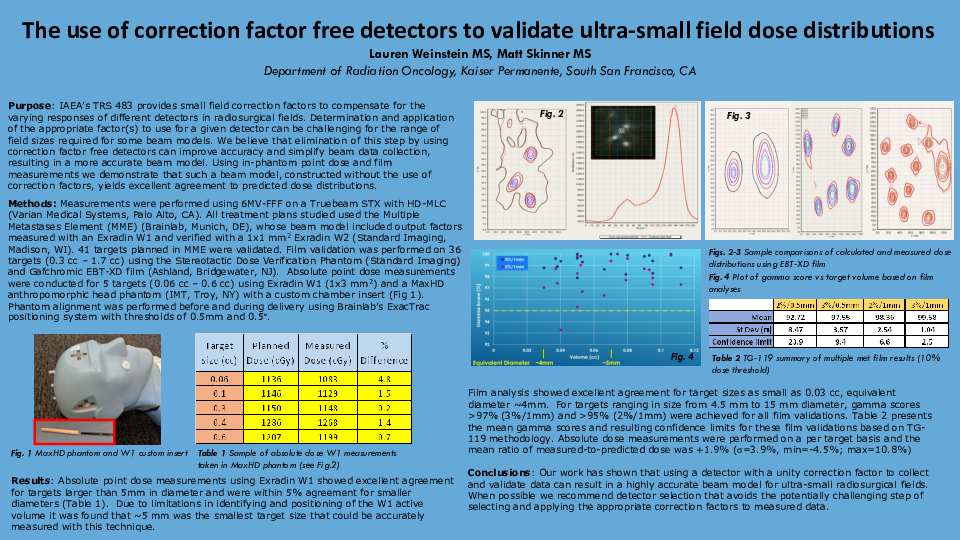
Purpose: Ultra-small field dosimetry (<1cm) is challenging due to perturbation effects and volume averaging. Recently, small field correction factors have been introduced to compensate for the the varying responses of different detectors in radiosurgical fields. We demonstrate that data acquired and validated with detectors not requiring correction factors, such as the W1 (Standard Imaging, Madison, WI) and Gafchromic film (Ashland, Bridgewater, NJ), yields excellent agreement between plans created in the Multiple Met (MME) Element (Brainlab, Munich, Germany) and phantom measurements.
Methods: Output factors were acquired on a Truebeam STX (Varian Medical Systems, Palo Alto, CA) with a flattening filter free 6 MV beam using an Exradin W1 scintillator. Treatment plans for 41 targets were validated in MME: 5 with the W1 and 36 with Gafchromic EBT3 or XD film. Target sizes ranged from 0.06 cc to 0.6 cc and 0.03 cc to 1.7 cc for the W1 and film validations, respectively. Treatment plans were planned and delivered on MaxHD (IMT, Troy, NY) and Baby Blue (Standard Imaging, Madison, WI) phantoms. Phantom alignment was performed before and during delivery using Brainlab’s ExacTrac with thresholds of 0.5mm and 0.5°.
Results: All plans validated with film and the W1 had good agreement with calculations. Gamma scores >96% (2%/1mm, 10% dose threshold) were achieved for all film validations for targets ranging from 0.03cc to 1.7cc. W1 point dose measurements were <1.5% of calculation for all but the 0.06cc target, at which the target size is comparable to the W1 dimensions.
Conclusions: Using a detector with a unity correction factor (or applying proper correction factors to an appropriate detector, such as those available in IAEA’s TRS 483) to collect and validate data is essential for an accurate machine build.


